Special projects between Bavaria and Florida
Lehrstuhl für Medizinische Physik
Friedrich-Alexander-Universität Erlangen-Nürnberg
Mathematics Department
University of Central Florida
Local Tomography for Cardiac CT
Cardiac and, more generally, dynamic imaging is a very important challenge facing modern computed tomography (CT). We plan to use local tomography to perform image reconstruction from motion contaminated data because it does not require irradiating parts of the object outside the heart. This property of LT is of even greater importance in cases when repeated scans are needed and were patient dose becomes a critical issue when ussing conventional approaches. We want to investigate how LT works on real clinical data and determine if it is effective in locating plaque in arteries.
Final report
Cardiac and, more generally, dynamic imaging is a very important challenge facing modern computed tomography (CT). The main difficulty is that the object being scanned changes during data acquisition. Hence the classical tomographic reconstruction theory does not apply. In a recent paper of the co-PI Dr. Katsevich it was shown how to perform image reconstruction from motion contaminated data using local tomography (LT). LT is a technique, which recovers a gradient-like image (i.e., with edges enhanced) of an object from its CT data. The main advantage of LT is that unlike conventional CT methods, it does not require irradiating parts of the object outside the region of interest (e.g., outside the heart in the case of cardiac imaging), thereby reducing the x-ray dose to the patient. This property of LT is of great importance in cases when repeated scans are needed, since even a single CT scan delivers significant x-ray dose to the patient. Another advantage of LT is its flexibility with respect to noise, motion, and other imperfections in the data. Cardiac imaging is one possible practical application of LT. For example, finding edges of blood vessels can be important for the diagnosis of the stenosis of the coronary arteries. An irregular-shaped narrowing of a blood vessel would indicate the presence of plaque. In the paper mentioned above LT was tested on simulated data.
In this project we investigated how LT works on real clinical data. The Institute of Medical Physics in Erlangen provided Prof. Katsevich with clinical data which he reconstructed with his algorithm. The data itself were acquired with a state of the art Siemens Somatom Definition Flash cone-beam spiral CT scanner during a clinical examination. This scanner has an interesting specialty, it has two acquisition systems orthogonal to each other. This increases the temporal resolution but also shows new possibilities of reducing the dose. The second system has a limited detector coverage and therefore a lower dose to not imaged areas. Up to now the full detector of the first system is needed for signal processing during the reconstruction. By using the reconstruction with LT these rays are no longer needed and so there is no need to measure them.
The dataset presented here was acquired with a sequence scan using prospective cardiac gating and a contrast agent. During the scan with 100 kV and a gantry rotation time of 300 ms the patient had an average heart rate of 59 bpm. The total time for this scan was 6.3 s, in this time four consecutive single circle scans were acquired with a total amount of 408 mAs. Both acquisition systems were used during acquisition, but only one system was used for reconstruction during this study.
Below we present the same slice reconstructed with two algorithms: Standard, is shown on the left. The second, based on local tomography, is shown on the right.
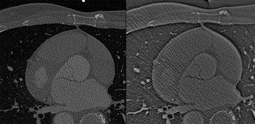
Comparing the images we see that the geometric features are all retained in the LT image. We thought this can be obtained with a significant reduction of the x-ray dose making LT a good tool for screening risk patients (for e.g. after implanting a stent to identify a restenosis or if there are risk factors within the family) more often than today. We expect that performing gated reconstruction of both acquisition systems will help to further increase the temporal resolution and the image quality at the moving heart.
Our results show that LT is a valuable tool if we thought of acquiring images with less dose as nowadays. This would enable us to monitor the health of patients with high risks at a much lower cost. For evaluating the value for the patient and the physician further research is required to fully investigate its potential for medical diagnostics.